The overarching goal of the Musculoskeletal Imaging and Orthopaedic Biomechanics (MIOB) Laboratory is to improve human health and performance for those who suffer from musculoskeletal and orthopaedic conditions. Our approaches span experimental and computational methods. We use magnetic resonance imaging (MRI) to visualize disease and quantify anatomy, and we image bone motion directly with dynamic x-ray. Our computational studies combine patient-specific MR-based models with dynamic motion measurements to estimate internal tissue mechanics of orthopaedic tissues. Overall, the experimental and computational methods employed by the MIOB lab aim to improve health outcomes and reduce burdens imposed on patients and our healthcare system.
Assistant Professor Department of Mechanical Engineering, Department of Electrical & Biomedical Engineering
Graduate Research Assistant: Mechanical Engineering
Arthokinomatic and qMRI changes of patients following combined ACL reconstruction and Meniscus surgeries
Motion capture, dual fluroscopy and MRI study
Graduate Research Assistant: Biomedical Engineering
Undergraduate Research Assistant: Biomedical Engineering
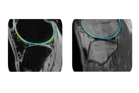
Biomedical Imaging of Musculoskeletal Tissue Structure, Size and Composition
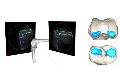
Dynamic Imaging of Joint Motion Using High-speed Dual Fluoroscopy
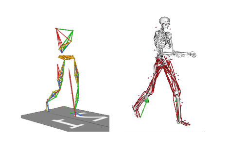
Joint Mechanics Estimation via Patient-Specific Computer Models