Noran Laser Scanning Confocal Microscope: Description and Capabilities
 This confocal microscope is optimized for live-cell or tissue imaging of fast calcium events measured using a green indicator such as fluo-3 or fluo-4.
- 50 mW Ar++ (488 nm) excitation
- Nikon inverted microscope Eclipse TE2000-U
- 500 nm long-pass emission filter
- Maximum imaging rate of 1 image in 2 ms with even faster data collection in linescan mode
- Complete electrophysiology setup with optical and electronic triggers for synchronization
- System was rebuilt by Prairie Technologies in 2005 and runs with PrairieView. Files are saved in standard TIFF format.
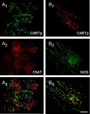
Left image: 3-D reconstruction of cardiac gangila with immunolabelling. A1, B1: CARTp immunoreactivity with FITC- and CY3-conjugated secondary antibodies. A2: ChAT-specific staining, B2: NOS-specific staining. A3, B3: Combined images that have been thresholded to highlight staining overlap. (ChAT= choline acetyltransferase, CARTp= cocaine- and amphetamine-regulated transcript peptide, NOS= nitric oxide synthase)
Calupca, et al., 2001, Journal of Comparative Neurology, 439:73-86

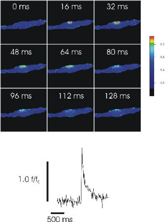
Right image: Cells loaded with fluo-3 and imaged at a rate of one image per 16 ms, viewed in pseudocolor. Bottom, F/Fo trace of fluorescence within a 2.2*2.2 micron area centered on the spark site. Excitation was at 488 nm with a 520 nm long-pass filter.
Wellman, et al., 2002, Stroke, 33:802-808